Category Archives: Optic Nerve

Most Glaucoma Patients are Undiagnosed

Glaucoma Awareness – Early Testing and Detection
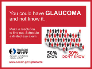
You could have Glaucoma and not know it Glaucoma has no symptoms and therefore it is often referred to as the sneak thief of sight. Patients with glaucoma experience no symptoms until the very late stages of the condition. Late stage glaucoma is characterized by profound vision loss.

Glaucoma Prevention

I am often asked by patients “I have a positive family history. How can I prevent myself from getting glaucoma?” The answer is early detection. We do this by being properly screened during your eye exam.

During your eye exam take advantage of the Comprehensive Digital Retinal Exam technology which allows us to take high-resolution panoramic and ultrasound images of the eye. These technologies combine hundreds of high-resolution images giving us a 3-dimensional view of the optic nerve. Some retinal nerve cells are more sensitive to glaucomatous changes than normal retinal cells. These retinal cells are called ganglion cells and when it comes to glaucoma they are kind of like the canaries in the coal mine. Ganglion cells are usually the first to signal early glaucomatous changes.

Yearly Eye Exams Reduce Blindness

By having yearly eye exams and comparing these digital images over the coming years, we look for subtle changes. Eye doctors usually diagnose glaucoma by noticing these subtle changes in the optic nerve over time.

Learning More

  1. There are two theories on glaucomatous vision loss, the vascular theory and mechanical theory.
  2. Some relatively new research also makes a compelling case that glaucomatous changes originate in the brain.
  3. Take the Glaucoma Eye-Q Test

Early Detection

Glaucoma ScreeningsIf you would like to start the new year off right we would be happy to asses your glaucoma risk factors at your next eye exam. Please call our Colleyville office at 817.416.0333 or the Keller office at 817.431.4900. You can also schedule your appointment online at any time.

Diopsys Pattern Electroretinograph Predicts Early Glaucomatous Vision Loss

Pattern Electroretinography (pERG) Gives up to 8 Years of Advanced Warning

Pattern ElectroretinographIncorporating the most advanced technology available in caring for our patients is important to the way we practice at Total Eye Care. The Diopsys® Pattern Electroretinograph is the newest addition to our suite of technology. The Pattern ERG (PERG) measures the signal strength of the retinal information being sent to the optic nerve. Studies show that a 10% decrease in this signal can be detected up to 8 years earlier with a Pattern Electroretinograph than a scanning laser ophthalmoscope.  This technology thus allows for much earlier diagnosis and treatment.

The Pattern Electroretinograph Helps Prevent Vision Loss in Glaucoma

Early detection and treatment of glaucoma is important due to the fact that nerve tissue cannot be regenerated. Twenty years ago, in the absence of a very high intra-ocular pressure, it was a common practice to wait until a visual field defect was present before initiating treatment. There is a significant amount of redundancy in the retinal nerve fibers. Studies have shown a patient must lose approximately 50% of the nerve fibers before visual field loss is detectable. The scanning laser ophthalmoscopes changed all of that allowing us to detect earlier, more subtle changes in the retinal nerve fiber layer. Now patients are treated before visual field defects become apparent.

The Diopsys® Pattern Electroretinograph is the next step in early detection. The PERG will not replace the scanning laser ophthalmoscopes such as the Zeiss Cirrus Optical Coherence Tomographer (OCT) in the diagnosis of glaucoma; however, it gives us another valuable tool in the diagnosis and treatment of glaucoma.

Visual Evoked Potential – New Technology Available at Total Eye Care

We have added a new service here at Total Eye Care. It is called the Visual Evoked Potential or VEP. Until recently this technology was only available to large institutions and teach hospitals. We are now able to provide this service for our patients at the Colleyville office.

What is the VEP?

The VEP, or Visual Evoked Potential, measures how well you visual system is working. Testing the integrity of your visual system is done by showing you a series of checkerboard patterns and then measuring the electrical activity between your eye and the visual cortex of your brain.

Your eye converts what you see into very low voltage electrical signals that travel along the optic nerve between your eye and the visual cortex. The computer inside the VEP compares the strength and speed of signal to a database of normal results and then the doctor uses that information to guide his or her diagnosis.

How is the VEP Done?

Two sticky pieces of tape are placed on your forehead and another on the back of your head. A patch is then placed over one eye and you look at a computer monitor that shows you the checkerboard pattern. When the testing is complete the process is reversed for the other eye. That’s all there is to it.

What Does the VEP Tell the Doctor?

The VEP evaluates how well the entire visual system is working. A visual field (peripheral vision test) gives the doctor similar information. In this way the VEP  helps the doctor diagnose and manage patients with glaucoma. The VEP is also used in the diagnosis and treatment of patients with amblyopia, multiple sclerosis, myasthenia gravis, optic atrophy, and concussion to name a few.

Scanning Laser Technology Available for Patients With Glaucoma & Macular Degeneration

New technology now available at Total Eye Care allows us to scan a patient’s retina for glaucoma and macular degeneration, and the best part ….. it does it  WITHOUT DILATION! This new instrument is called the Zeiss Cirrus OCT and it is truly state of the art.

The Zeiss OCT uses infra-red light from a scanning laser to make a very high resolution, 3D image of the inside of your eye, much like that of an MRI. We can now identify retinal objects as small as 4 microns. So how small is 4 microns? A single sheet of 20 lb copy paper is 100 microns thick. This new technology allows us to detect glaucoma and macular degeneration much earlier as well as allowing us to detect and track subtle retinal changes.

Who is a candidate for this new technology? Anyone with a family or personal history of glaucoma, glaucoma suspect or macular degeneration should take advantage of this technology.

Call the Colleyville office at 817.416.0333 and let Rona or Kelsey know that you would like to take advantage of the new scanning laser technology right away.

Brain May Be The Key To Early Glaucoma Detection

David Calkins, Ph.D., Director of Research at the Vanderbilt Eye Institute published a study indicating that the brain may hold the key to the early signs of glaucoma.  Published in the Proceedings of the National Academy of Sciences, the study indicates that glaucoma may be like other central nervous system disorders with its origins being in the brain.

Traditional thinking regarding glaucoma is that the either the high pressure slowly crushes the nerve fibers or the pressure decreases blood flow to the optic nerve.  Should Dr. Calkins’ findings be confirmed in human studies it would cause a paradigm shift in the treatment and diagnosis of glaucoma.  Dr. Calkin’s study demonstrates that glaucoma starts in the brain and then as the disease progresses the optic nerve starts to show evidence of the disease.  Currently Dr. Calkins’ research is directed toward looking for medical therapies that can restore the connection of the nerve fibers between the brain and the retina.

I have to applaud Dr. Calkins and his team for thinking outside the box in their pursuit of answers to the second leading cause of blindness.  The study was funded by the National Eye Institute, the Glaucoma Research foundation and Research to Prevent Blindness.  More details on the study results are available on The Reporter, Vanderbilt Medical Center’s Weekly Newspaper.

World Glaucoma Awareness Week is March 7 to March 13

FOR IMMEDIATE RELEASE

COLLEYVILLE, TEXAS – March 6, 2010 – In honor of World Glaucoma Awareness Week (March 7 to March 13, 2010), Total Eye Care will be offering free Glaucoma Screenings on World Glaucoma Day, March 12, 2010 at the Colleyville office located at 6114 Colleyville Blvd. from Noon to 2pm.

“Early detection and awareness are crucial to limiting vision loss from glaucoma.  The purpose of Glaucoma Awareness Week is to get the word out and prompt patients to seek an evaluation from their eye doctor.  At Total Eye Care we would like to make it easier for patients to learn about glaucoma by offering a free screening ” says Dr. Richard Driscoll, clinical director at Total Eye Care.  Glaucoma is the second leading cause of blindness worldwide, not because of lack of effective methods for diagnosis and treatment, but due to insufficient information available to the public and low awareness of the implications of the disease.  Glaucoma is asymptomatic in its early stages, while the patient’s vision suffers irreversible and cumulative damage. Visual disturbances are noted by the patient during the later stages on the way to blindness, whereas timely diagnosis and therapy could have prevented this degradation.

“We have so many advanced medications and diagnostic instruments available to us that the detection and treatment of glaucoma has been greatly advanced in the last 10 to 15 years” says Dr. Alycia Green.  A glaucoma awareness questionnaire is available on the Total Eye Care website along with detailed handouts for patients on glaucoma and many other eye diseases such as macular degeneration and keratoconus.

Total Eye Care participates in Glaucoma Awareness Week, the complete press release.

About Total Eye Care

Therapeutic Optometrist and Optometric Glaucoma Specialist Dr. Richard Driscoll has been serving the eye care needs of Colleyville, Texas since 1995.  Dr. Alycia Green brings over 10 years of experience to Total Eye Care, specializing in pediatrics.  The Colleyville office is located at 6114 Colleyville Blvd.  Total Eye Care’s Keller office is located at 1834 Keller Parkway. More information can be found on The Eye Doc Blog or the Total Eye Care website at www.TotalEyeCare.com

Contact Information

Dr. Richard Driscoll
Total Eye Care
6114 Colleyville Blvd.
Colleyville, TX 76034
817.416.0333

Pilot Study* Indicates Targeted Ophthalmoscopy With the Optomap® Retinal Exam Increases Ocular Disease Detection

Results from a pilot study conducted by doctors at the New England College of Optometry were recently presented at Optometry’s Meeting and the American Academy of Optometry.  In the study, subjects were examined using the methods of traditional ophthalmoscopy and optomap® assisted ophthalmoscopy, where an ultra-widefield retinal image from the Optos P200C guides the BIO fundus examination.

Forty subjects underwent non-mydriatic imaging with the P200C. Subjects were then dilated and underwent traditional ophthalmoscopy and optomap® assisted ophthalmoscopy. The traditional exam consisted of BIO and slit lamp biomicrosopy with precorneal lenses. optomap® assisted ophthalmoscopy was identical to traditional, with the exception that the doctor reviewed optomap® Images before performing their BIO examination. A masked Reader graded the optomap® Images of all subjects. A retinal specialist examined 28 of the 40 subjects and served as the gold standard. Continue reading

The Connection Between Light and Migraines

It seems that virtually everyone that has experienced a migraine wants to crawl into a dark room.  The February 2010 issue of the journal Nature Neuroscience has published a study that explains why this phenomena may occur.

The study links a possible connection between light sensitive retinal nerve cells and nerve cells in the brain that are responsible for the perception of migraine pain.  The study noted that blind patients also avoided light when suffering from a migraine, however blind patients that had lost their eye did not avoid light.  The authors concluded that some of the retinal axons, included some light sensitive axons were sending signals to the hypothalamus, a part of the brain that is responsible for the perception of headache pain.

FDA Nears Approval of Eyelash Lengthening Drug

Allergan, Inc. the maker of Lumigan, a medication we currently use to lower the intra-ocular pressure in patients with

Photo, Lumigan ophthalmic solution

Photo, Lumigan ophthalmic solution

glaucoma, is expected to receive approval to market Lumigan to patients that wish to lengthen and thicken their eyelashes.  Lumigan is a popular drug for the treatment of glaucoma.  A side affect of Lumigan is that of thickening and lengthening the eyelashes.  Patients often ask us if there is a way to make their eyelases longer and thicker.  The answer was always yes but it’s not approved for that purpose.

The “new drug” will be called Latisse (Bimatoprost Solution 0.03%) and is pending approval for the treatment of hypotrichosis of the eyelashes (reduced amount of hair).  Latisse will cause an increase in the number, thickness and darkness of the eyelashes.  Unlike Lumigan it is not intended to go directly into the eye, but rather on the eyelashes.  Allergan expects to begin marketing Latisse in 2009.

As a side note a few years ago a patient brought a product from the Jan Marini line of skin care products into the office.  The company claimed the product contained a drug that caused the eyelashes to lengthen.  Not long after that the FDA forced it off the market.  I’m glad to see an alternative is just around the corner.

The FDA approves LATISSE(TM) December 26, 2008, read about it here.

How Does Glaucoma Damage the Eye?

Glaucoma Blindness Theories

We are frequently asked to explain how glaucoma causes blindness. Glaucomatous damage to the eye is caused when the pressure within the eye is greater than the optic nerve can tolerate. How does the pressure cause blindness? The simple answer is we don’t know exactly; however, there are two main theories as to why the damage occurs. Neither theory fully explains how the optic nerve damage occurs in the different types of glaucoma.

The Vascular Theory of Glaucoma

The premise of the vascular theory is that high pressure inside the eye restricts blood flow to the optic nerve causing the optic nerve to slowly die from lack of oxygen and nutrients.

The Mechanical Theory of Glaucoma

Physical glaucoma damage is the underlying hypothesis of the mechanical theory of glaucoma. It is thought that the high pressure damages the optic nerve fibers.

So Which One Is It?

That’s a good question the leading researchers can make a convincing case for both theories and neither theory, by itself, completely explains how glaucoma damages the eye. In reality, both probably play a role in how an eye is damaged by glaucoma.

Stumble It!